What Is Retinal Ischemia and Why Does Blood Flow Matter?
Like every tissue in the body, the retina relies on consistent blood flow to deliver the oxygen and nutrients it needs to function properly. When blood flow is reduced or interrupted, retinal cells can become stressed, damaged, and eventually die. This is known as retinal ischemia, a complication seen in several common conditions, including diabetes, retinal vein occlusions, high blood pressure, and carotid artery disease. Left untreated, retinal ischemia can lead to permanent vision loss. Here's a closer look at the conditions that can disrupt blood flow to the retina and why healthy circulation is essential for preserving vision.
What Is Retinal Ischemia?
The retina is a light-sensitive layer of tissue located at the back of the eye. It plays a critical role in vision by converting light into signals that are interpreted by the brain and translated into the images we see. Like other highly active tissues in the body, the retina requires significant amounts of oxygen, which depends on a healthy network of blood vessels to deliver a steady supply of oxygen-rich blood. Retinal ischemia occurs when these blood vessels become damaged or blocked, and the retina does not receive adequate blood flow.
Conditions That Can Cause Retinal Ischemia
Several common conditions can interfere with circulation and increase the risk of retinal ischemia.
Diabetes
In those with diabetes, elevated blood sugar levels can damage the small blood vessels that supply the retina. When this occurs, these vessels can swell, leak fluid, or close off entirely, which restricts blood flow to the retina. In earlier stages, this is known as non-proliferative diabetic retinopathy, which can cause swelling in the macula, the part of the retina responsible for central vision. As the disease progresses, the retina may begin growing new, abnormal blood vessels in an attempt to compensate for lost circulation. These fragile vessels are prone to bleeding and can cause significant vision problems if not treated.
Retinal Vein Occlusion
Retinal vein occlusion occurs when one of the veins draining blood away from the retina becomes partially or fully blocked. Without a clear path out, blood and fluid back up into the retina, causing swelling and elevated eye pressure that prevents oxygen-rich blood from entering the retina. There are two main types of retinal vein occlusion. Central retinal vein occlusion involves the main drainage vein, while branch retinal vein occlusion affects one of the smaller veins. Both can lead to vision loss and require prompt evaluation.
High Blood Pressure
Long-term high blood pressure places excess force on blood vessel walls throughout the body, including the small vessels in the retina. Over time, this can cause those vessels to narrow, thicken, or leak, reducing the retina's blood supply and impairing its ability to function. This is known as hypertensive retinopathy. Many patients with hypertensive retinopathy do not notice symptoms until the damage is already substantial.
Carotid Artery Disease
The carotid arteries supply oxygen-rich blood to the head and neck, and therefore the eyes. When fatty deposits build up and narrow these arteries, blood flow can become blocked. In some cases, a piece of plaque can break off and travel to the arteries that supply blood to the retina, causing a sudden blockage. This is known as a retinal artery occlusion, which is essentially a stroke of the eye. A retinal artery occlusion can result in sudden vision loss and must be evaluated immediately.
Symptoms of Retinal Ischemia
Symptoms can vary depending on which part of the retina is affected and how severely blood flow is reduced. Some patients notice symptoms suddenly, while others experience more gradual changes.
Potential symptoms include:
- Blurry or distorted vision
- Dark or missing areas in vision
- Reduced peripheral vision
- Difficulty seeing fine details
- Partial or sudden vision loss
Any sudden vision change should be treated as a medical emergency.
How Retina Specialists Diagnose Retinal Ischemia
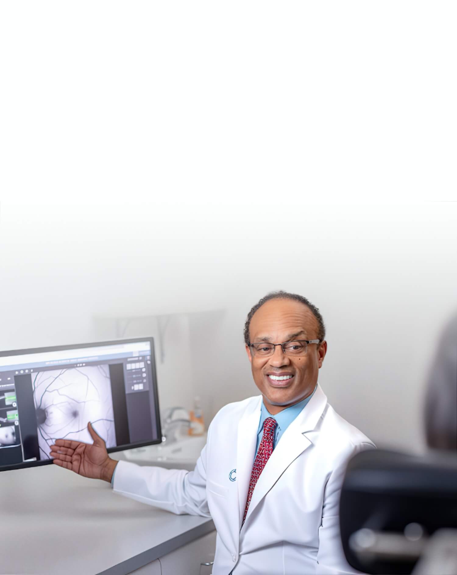
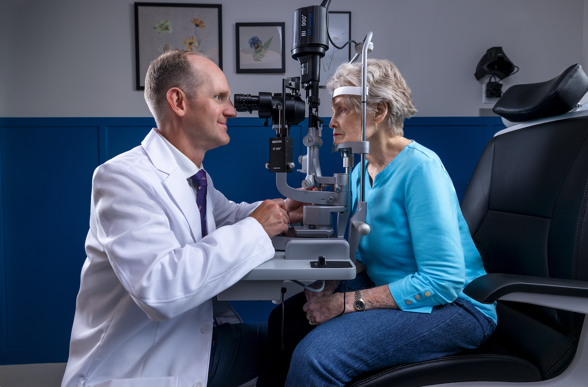
Diagnosing retinal ischemia begins with a comprehensive dilated eye examination, which allows retina specialists to see signs of vessel damage, swelling, or abnormal growth in the retina. For further evaluation, your provider may also use optical coherence tomography (OCT) to get more detailed images of the retinal tissue and get a clearer picture of any swelling and/or structural changes. In some cases, your provider may also recommend fluorescein angiography, which is an imaging procedure that uses a specialized dye to track blood flow through the retinal vessels and identify areas where circulation has been compromised.
Managing Retinal Ischemia
Managing the underlying conditions of retinal ischemia, such as diabetes, high blood pressure,, and cardiovascular disease, plays an important role in preserving retinal circulation and reducing the risk of further damage. Therefore, working closely with your primary care physician or specialist, alongside your retina specialist, gives your eyes the best chance of staying healthy long term.
If you have been diagnosed with diabetes, high blood pressure, or another condition associated with retinal ischemia, the team at Retina Consultants of Texas is here to help. Early detection and consistent monitoring make a meaningful difference in outcomes. To learn more, reach out or request an appointment today.